Dental X-ray: panoramic X-ray and 3D scan (CBCT) in Baku
Dental X-rays show what the eye cannot see: decay between the teeth, inflammation at the tip of a root, the shape of the canals. One is taken only when it is needed.
25–95 AZN, 1 scanThe exact price is clear after the examination.

Does this sound familiar?
- My tooth hurts, but the cause cannot be seen
- I need a scan before root canal treatment
- I want to get braces or aligners
- My tooth was injured and I want it checked
- There may be a cyst at the root of my tooth
- I have an older X-ray and would like you to look at it
An image is taken only when it is needed, and you are told which one and why.
What does a dental X-ray show?
Diagram: a flat X-ray and a 3D scan
- A flat X-ray shows the tooth, the roots and the bone on one plane, so they can overlap.
- A 3D scan (CBCT) shows the same area as a volume.
- It can be viewed in any slice: across the roots, each canal shows separately.
A dental X-ray shows the inside of the tooth, the roots and the bone. The right treatment often starts with the right image. There are three kinds of dental X-ray. The clinic has a panoramic X-ray and a 3D scanner (CBCT), so both are taken on the spot.
- Small X-ray Shows one or two teeth close up. If you had one taken elsewhere, bring it.
- Panoramic X-ray All the teeth and both jaws in one flat image. It shows the overall state, how a child's teeth are developing, where the wisdom teeth are. It is taken at the clinic.
- 3D scan (CBCT) Shows the tooth, the roots and the bone in three dimensions. Needed for complex root canal treatment and to see the size of a cyst and the bone precisely. It is taken at the clinic.
How does an X-ray examination go?
Examination
First the tooth is examined and your concerns are heard. If an image is needed, you are told which one and why.
Older images
If you have earlier images, they are looked at first. Sometimes a new one is not needed.
Taking the image
Both the panoramic X-ray and the 3D scan are taken right at the clinic. You wear a protective apron when needed.
Treatment and checks
The treatment is planned from the image, and you are shown what it reveals.
When you come in
- If you have earlier images, bring them with you.
- If you are pregnant, always tell the dentist.
What does the price of a dental X-ray depend on?
A panoramic X-ray is 25 AZN. A 3D scan: half a jaw 30 AZN, one jaw 55 AZN, both jaws (upper and lower) 95 AZN, the jaw joint 50 AZN. Both are taken at the clinic. Whether you need one, and which, is decided at the examination.
What changes the price
- The kind of image: a panoramic X-ray or a 3D scan
- The area of a 3D scan: half a jaw, one jaw, both jaws or the jaw joint
- If you have an earlier image, a new one may not be needed
Frequently asked questions
Are dental X-rays harmful?
The radiation from modern dental X-rays is very low. Even so, an image is taken only when it is needed, with a protective apron where needed. A 3D scan gives more radiation than a normal X-ray, so one is taken only when it is really necessary.
How much does a dental X-ray cost?
A panoramic X-ray is 25 AZN. A 3D scan: half a jaw 30 AZN, one jaw 55 AZN, both jaws (upper and lower) 95 AZN, the jaw joint 50 AZN. Whether you need one is decided at the examination. If you have an earlier image, bring it: sometimes a new one is not needed.
What is the difference between an X-ray and a 3D scan?
A normal X-ray shows the tooth flat, as a two-dimensional picture. A 3D scan shows the tooth, the roots and the bone in three dimensions: you can see how many canals there are, their shape, and the real size of a cyst. The clinic takes both: a panoramic X-ray for the overview, and a 3D scan only when it is needed.
When is a 3D scan needed?
When the inside of the tooth, the roots and the bone need to be seen precisely. For example, in complex root canal treatment, to see how many canals there are and their shape, or to find out the real size of a cyst. It gives more radiation than a normal X-ray, so one is taken only when it is really necessary.
Can I have a dental X-ray while pregnant?
The radiation from modern dental X-rays is very low, but planned X-rays wait until after the birth. If urgent treatment is needed because of pain or inflammation and it cannot be done without an image, an image can be taken with a protective apron. Always say that you are pregnant.
Can I bring an older X-ray?
Yes. If you have earlier images, bring them to your visit. Sometimes looking at them is enough and a new one is not needed. If a new image is needed after all, you are told which one and why. An image is taken only when it is needed.
What is a panoramic dental X-ray?
A panoramic X-ray shows all the teeth and both jaws on one flat image: the overall picture, for example how a child's teeth are developing and where the wisdom teeth are. The clinic takes panoramic X-rays too. When a closer look is needed, a 3D scan is taken: it shows the area needed in three dimensions. Which image you need becomes clear after the examination.
Read in more detail
Panoramic X-ray and 3D scan: the difference in plain words
A panoramic X-ray is like an unfolded map of the mouth: all the teeth and both jaws on one flat picture. A 3D scan is more like a model: you can look at the same area from every side and in any slice.
The map gives the overall picture quickly, but details can overlap. The model is more precise, but it is only needed when there is a specific question. The clinic takes both: a panoramic X-ray for the overview, a 3D scan for a specific question.
- A panoramic X-ray shows well The state of all the teeth, teeth that have not come through yet, the roots in general, large areas of inflammation.
- A panoramic X-ray shows poorly Small decay between teeth, fine detail of a single tooth.
- A 3D scan shows well The number and curve of the canals, the thickness of the bone, the real size and position of a cyst.
Radiation compared with everyday life
Everyone receives natural radiation every day: from the ground, the air and food, and a little more when flying. Dental X-rays are among the lowest-radiation tests in medicine. A small or panoramic X-ray is comparable to the natural background a person receives over a few days of ordinary life.
A 3D scan gives more radiation than a normal X-ray, but usually much less than an ordinary CT scan of the head. That is why one is taken only when there is a specific question, not every time.
- An image is taken only if it will answer a question.
- The area imaged is kept as small as possible.
- A protective apron is used when needed.
- Your earlier images can mean you do not need another one.
Which image is needed when?
Each problem needs its own kind of image, and sometimes no image is needed at all. The clinic takes panoramic X-rays and 3D scans. If you have a small X-ray from another clinic, you can bring that too. The list below is the general rule. The final decision is made after the examination.
- Complex canals A 3D scan, to see how many canals there are and how they curve.
- Suspected root cyst or an injured tooth A 3D scan, to see the real size of the cyst, the root and the bone.
- Braces or aligners An overall picture of the teeth, roots and jawbone. At the clinic this is a panoramic X-ray or a 3D scan.
- A child's teeth developing or where the wisdom teeth are: a panoramic X-ray.
- Small decay between teeth It usually shows on a small X-ray. If you have one, bring it.
What happens while the image is taken?
It is quick and painless. The main thing is to keep still for those few seconds. If you move, the image is blurred and has to be retaken.
- For both a panoramic X-ray and a 3D scan, you rest your chin on a support and the machine moves around your head.
- Take off metal items beforehand, such as earrings, necklaces, glasses and hair clips.
- Ask for a digital copy of the image. It is useful to show another doctor and to compare later.
The dentist's courses in this field
- years of experience, since 2004
- 20+
- courses and conferences in this field
- 5

2nd International Digital Orthodontics conference of the Azerbaijan Orthodontic Society.

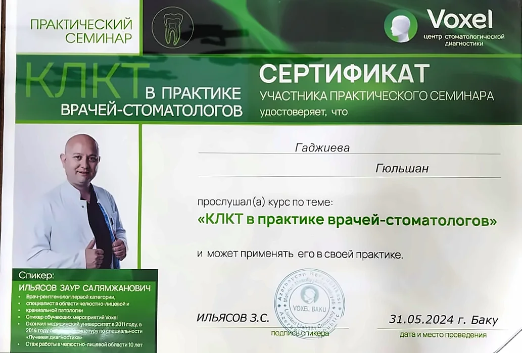
Hands-on seminar on cone beam CT (3D scans) in dental practice.
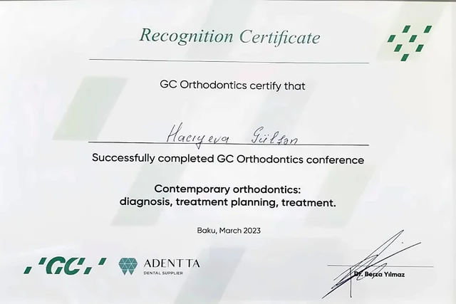
GC Orthodontics conference on diagnosis, treatment planning and treatment.

Course on digital cephalometry (orthodontic diagnosis).
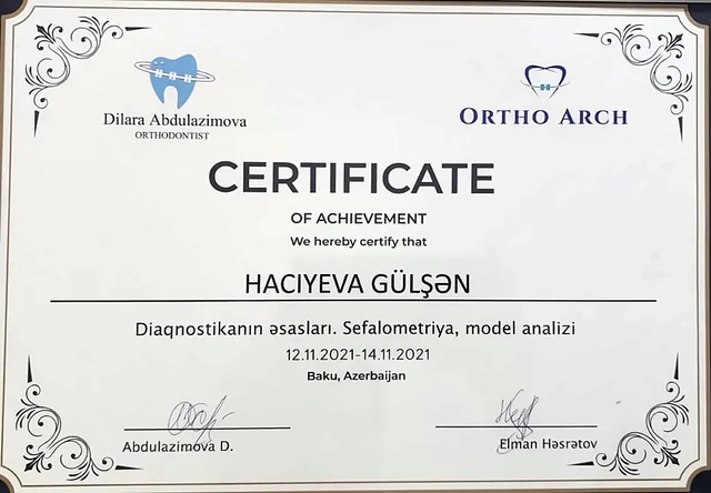
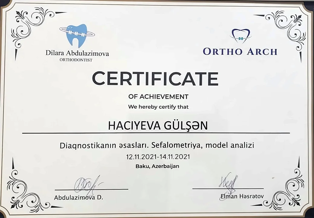
Basics of orthodontic diagnosis: cephalometry and model analysis.


- 2026
Certificate to practise
State certificate to practise, dentistry
- 2002
University
Azerbaijan Medical University named after N. Narimanov
Other services

Not sure whether you need an image?
Send a message on WhatsApp and briefly describe what bothers you. Which image you need is decided at the examination.